In recent years, sport conditioning practices have placed increasing emphasis on improving ankle range of motion (ROM), with forced dorsi flexion often being a key component. The idea is to alleviate tension in the Achilles tendon and calf muscles, enhancing mobility and performance. However, this approach tends to overlook the complex interplay of structures crossing the ankle joint—most notably the tibialis posterior muscle. Neglecting this key muscle’s anatomy and function can inadvertently increase injury risk and compromise performance.
Anatomy and Function of the Tibialis Posterior
The tibialis posterior is a long, spindle-shaped muscle located deep within the posterior compartment of the lower leg, sandwiched between the tibia and fibula. Its proximal attachments include the posterior surfaces of the tibia and fibula, as well as the interosseous membrane. Distally, the tendon inserts primarily on the navicular bone, with additional fibers extending to the cuneiforms, cuboid, and the bases of the second, third, and fourth metatarsals.
The tibialis posterior serves several critical roles:
- Stabilization of the medial arch: This muscle is a primary stabilizer of the foot's medial longitudinal arch, preventing overpronation.
- Inversion of the foot: It plays a key role in foot inversion, contributing to proper gait mechanics.
- Plantar flexion assistance: It aids the larger calf muscles in plantar flexion, especially during dynamic activities like running or jumping.
- Energy transfer during gait: It supports efficient energy transfer and shock absorption.
- Support under load: The muscle adapts to manage forces during weight-bearing activities, ensuring ankle stability.
Isometric Stabilization in High-Speed Running
During high-speed running, the tibialis posterior, flexor digitorum longus (FDL), flexor hallucis longus (FHL), peroneals, and the calf muscles (gastrocnemius, soleus, and plantaris) work near-isometrically to stabilize the foot. This coordinated effort is critical for managing shock absorption and maintaining the structural integrity of the foot.
The near-isometric behavior of these muscles during the stance phase helps the foot maintain a stable platform for force transfer while mitigating excessive joint motion. This stabilization allows the tendons to act as energy storage and release systems, reducing the metabolic cost of running and protecting the lower limb from excessive strain. Disrupting this intricate balance—such as by weakening or overstretching one or more of these stabilizing muscles—can lead to inefficiencies, reduced shock absorption capacity, and heightened injury risk.
The Problem with Forced Dorsi Flexion
In attempting to reduce tension in the calf muscles (gastrocnemius, soleus, and plantaris), forced dorsi flexion inadvertently reduces the functional capacity of the tibialis posterior and other synergists like the FDL and FHL. This redistribution of load places additional stress on the calf muscles and Achilles tendon, disrupting the natural synergy of these structures. Over time, the altered force dynamics can lead to compensatory mechanisms, tissue overuse, and microtrauma.
Worse yet, the implications extend beyond the tibialis posterior and calf complex. Forced dorsi flexion may overstretch the tendons of these muscles, diminishing their force production capabilities and increasing the risk of chronic injuries and surgery.
A Contributing Factor to Achilles Tendon Injuries?
The rise in Achilles tendon injuries among athletes may, in part, stem from this misguided focus on forced dorsi flexion. By prioritizing ROM gains without considering the holistic function of the ankle’s musculature, strength coaches risk compromising joint stability and overloading critical structures.
A More Balanced Approach
Rather than overemphasizing dorsi flexion, a more comprehensive strategy should:
- Include targeted strengthening of the tibialis posterior and associated muscles to enhance medial arch stability and near-isometric performance.
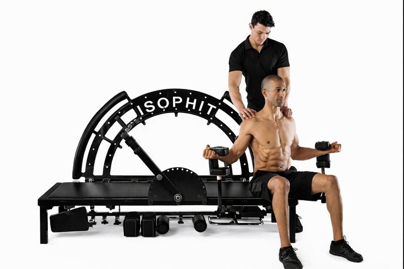
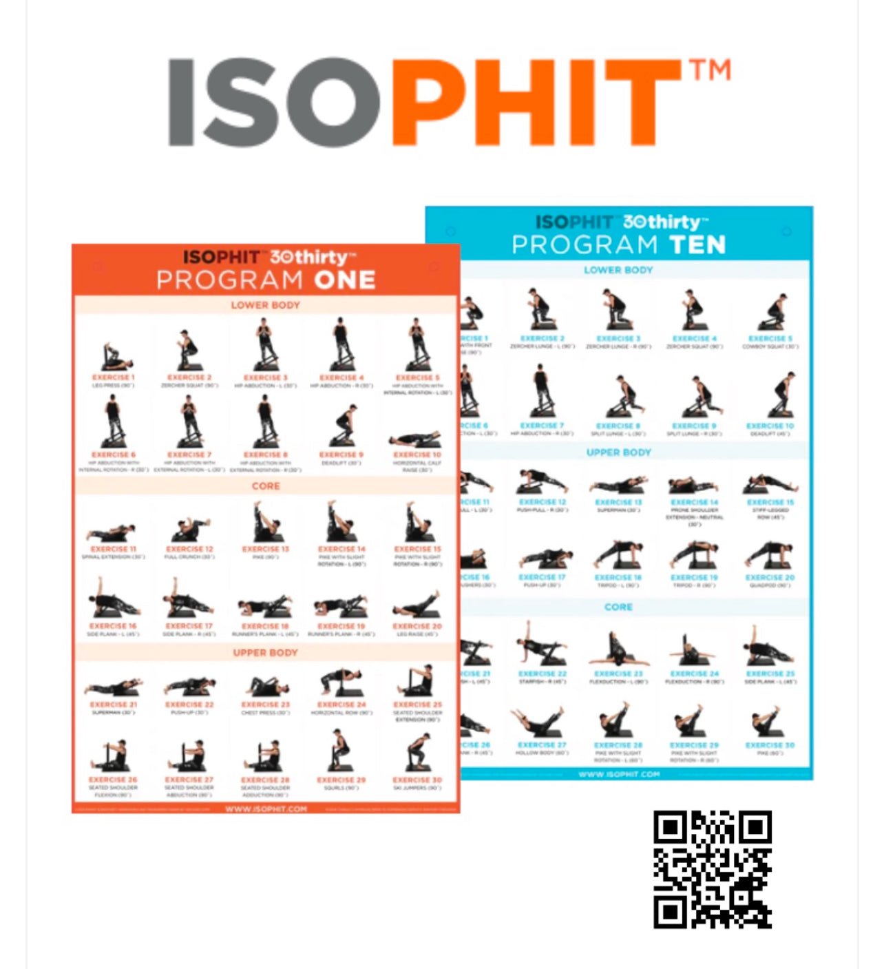
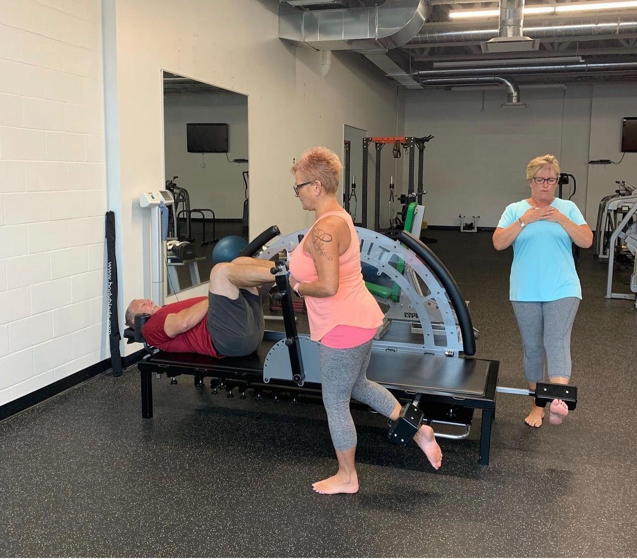
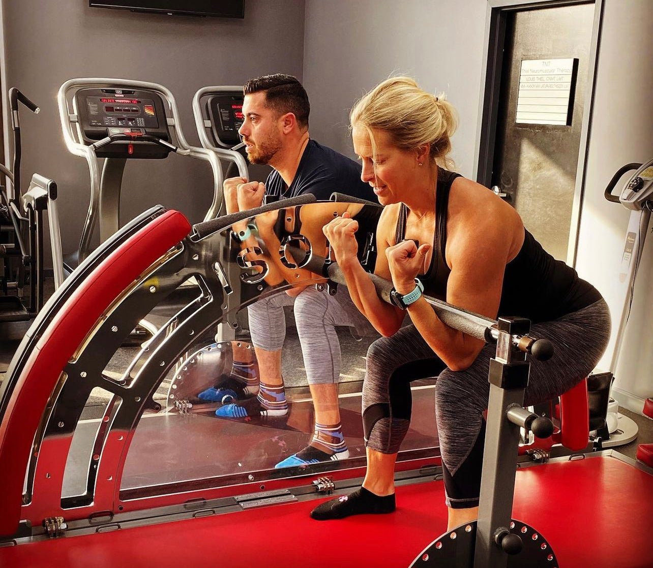
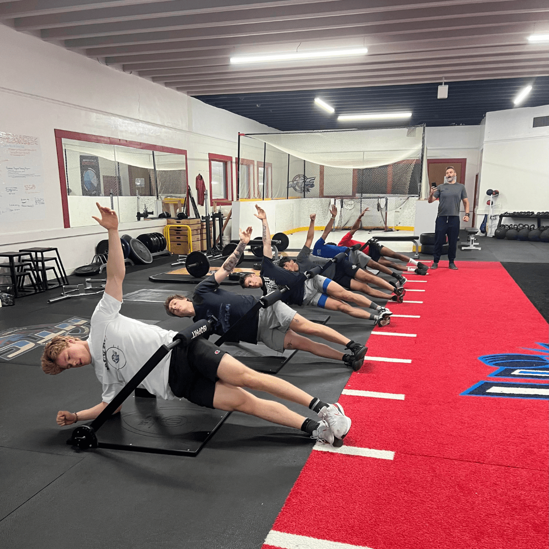
- Focus on additional isometric strength training drills that respect the natural biomechanics of the ankle joint. (see below)
- Incorporate whole-body isometric exercises that reinforce coordinated foot stabilization during high-speed and high-impact activities.
- Address compensations in related structures, such as the hip and knee, to reduce unnecessary stress on the lower leg.
Ultimately, a nuanced understanding of ankle anatomy and function is critical for minimizing injury risk while optimizing performance. Coaches and strength specialists must resist the allure of one-size-fits-all solutions and instead embrace evidence-based approaches that prioritize long-term athlete health.
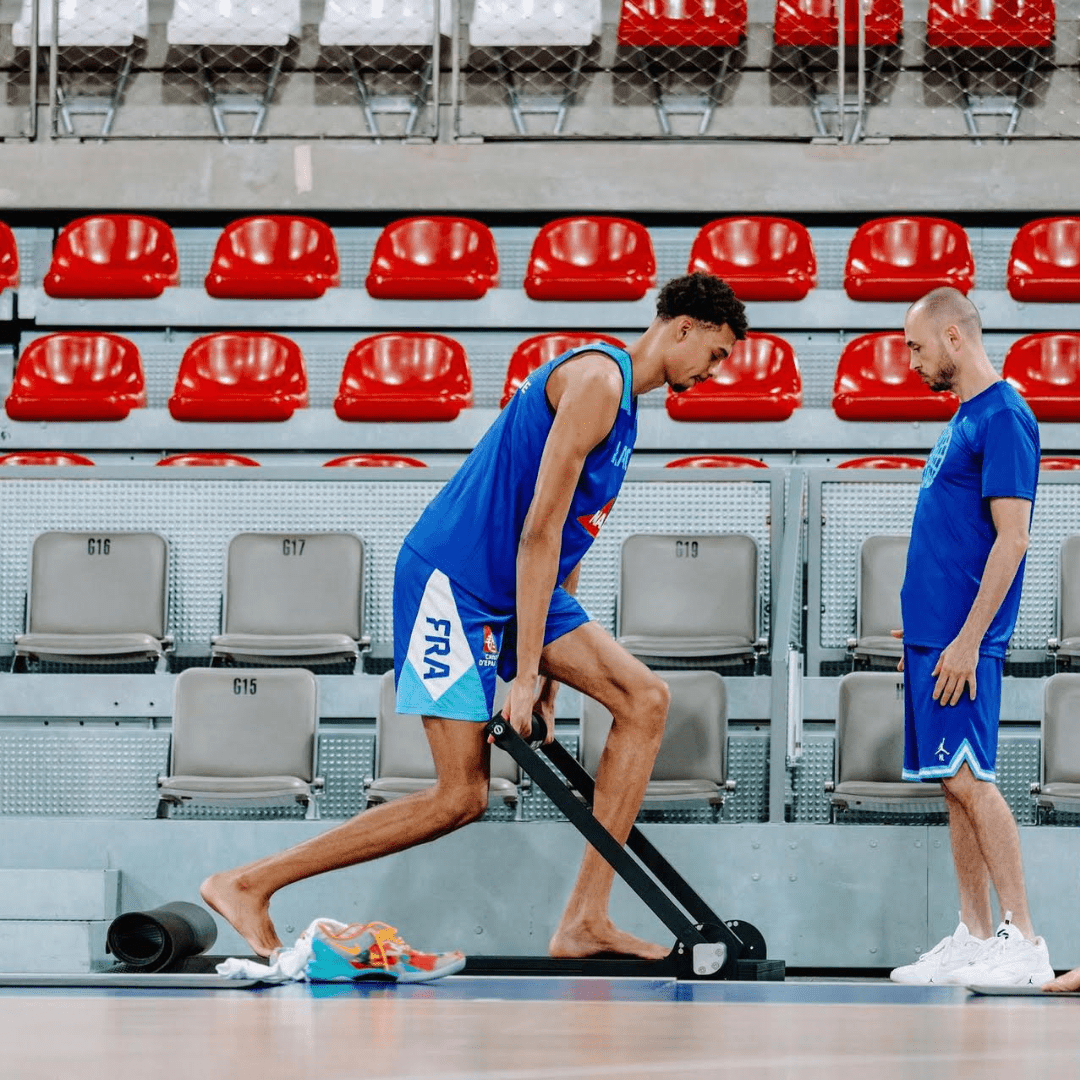
At Isophit, we help the world's strongest, fastest, and most gifted athletes win more, hurt less, and age stronger.


Share:
The Role of Isometric Strength in Power Production
Why Train Multiple Isometric Exercises?